Advertisement
)
AT A GLANCE
- Concept: Positional Context: The system reads genetic data without grinding the tissue into an unrecognizable chemical soup.
- Concept: Biochemical Barcoding: Millions of microscopic probes tag individual RNA molecules with unique geographic coordinates.
- Concept: Multiplexed Fluorescence: Lasers illuminate these tagged molecules to visually map cellular activity across a glass slide.
- Concept: Solid Tumors: Oncologists use this topographical map to see exactly how cancer cells evade surrounding immune systems.
HOW IT WORKS
Traditional genetic sequencing operates exactly like a blender. Scientists take a physical tissue sample, liquefy it to extract the genetic material, and run it through a standard sequencer.
This legacy method reveals exactly which genes are active within the sample. However, it completely destroys the physical architecture of the tissue, mixing millions of distinct cells into one average genetic signal.
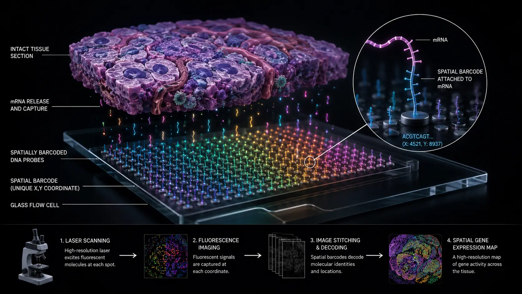
Spatial transcriptomics solves this destruction by treating a glass microscope slide as a high-density biological hard drive. Engineers print millions of microscopic spots onto this glass, with each spot containing billions of synthetic DNA capture probes.
Every single probe inside a specific spot carries an identical, unique spatial barcode. This microscopic ZIP code represents the exact physical coordinate of that specific spot on the glass slide.
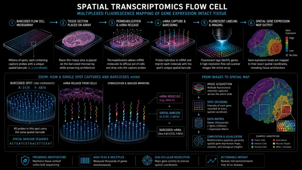
Technicians place a razor-thin slice of intact biological tissue directly on top of this barcoded grid. They apply a chemical solvent to permeabilize the cell membranes, causing the tissue’s messenger RNA to drop straight down onto the glass.
The synthetic probes instantly grab the falling RNA molecules and permanently attach the geographic barcode to them. A high-resolution flow cell laser then scans the slide, reading fluorescent chemical tags to map exactly which genes are expressing at every specific coordinate across the tissue geometry.
WHY IT MATTERS NOW
The global pharmaceutical industry currently wastes billions of dollars developing oncology drugs that fail in human trials.
Solid tumors do not exist as uniform masses of identical mutant cells. They operate as highly complex, fortified biological cities containing blood vessels, structural tissue, and actively suppressed immune cells.
Bulk sequencing cannot map these hostile microenvironments because it blends the tumor cells together with the immune cells, mathematically erasing the critical physical boundaries.
Spatial transcriptomics allows pathologists to see the exact border where a tumor chemically deactivates an approaching white blood cell. This topographical intelligence dictates the entire future of precision oncology.
Companies like 10x Genomics and Illumina race to dominate this hardware ecosystem, with Illumina officially launching its sequencing-based StrataMap Spatial Solution to directly challenge 10x’s market share.
For investors, the equipment required to generate this data represents an extreme capital moat. High-plex spatial imagers and the accompanying proprietary chemical reagent kits cost research hospitals hundreds of thousands of dollars per unit, securing highly lucrative, recurring revenue streams for the hardware manufacturers.
WHAT MOST PEOPLE MISS
Medical media assumes spatial biology is purely a hardware engineering challenge involving better lenses and brighter lasers. They completely miss the immense computational burden this technology creates.
Scanning a single one-centimeter tissue sample at subcellular resolution generates several terabytes of raw image data in a matter of hours. Standard hospitals lack the localized server infrastructure to process this sheer volume of information.
Storing, stitching, and analyzing these massive spatial datasets requires routing highly sensitive patient data through external cloud computing pipelines. The primary bottleneck blocking widespread clinical adoption is not physical chemistry; it is the absolute lack of hospital-grade data bandwidth and bioinformatics storage capacity.
THE TRAJECTORY
Next 12–36 Months: Pharmaceutical companies will mandate spatial transcriptomics profiling for all late-stage clinical oncology trials. This structural data will allow trial operators to retroactively identify exactly why a specific immunotherapy cured one patient but failed entirely in another.
Next Five Years: Multi-omic spatial imagers will commercialize heavily. These systems will simultaneously map messenger RNA, surface proteins, and cellular metabolites on a single slide, providing a complete, unbroken biochemical view of tissue function.
Next Ten Years: Spatial transcriptomics will move from the research laboratory into routine clinical diagnostics. Biopsy analysis will automate completely, utilizing machine learning algorithms to instantly grade tumor severity based on the spatial arrangement of malignant cells.
What Could Go Wrong: The chemical reagents used to permeabilize the tissue are highly sensitive to microscopic environmental changes. If hospital technicians deviate from the strict timing protocols by even a few seconds, the RNA degrades instantly, rendering the expensive spatial map completely blank.
Most Likely Outcome: Spatial biology will permanently replace bulk sequencing for all solid tissue analysis. The technology will transition from an expensive research novelty into the foundational diagnostic tool required to prescribe personalized genetic medicine.
KEY TERMS
- Spatial Transcriptomics: A molecular profiling technique that maps gene expression activity to exact physical locations within an intact tissue sample.
- Messenger RNA (mRNA): A single-stranded RNA molecule that carries the genetic instructions from a cell’s DNA to its protein-making machinery.
- Spatial Barcode: A unique, synthetic DNA sequence used to chemically tag molecules with their specific geographic coordinate on a microscope slide.
- Multiplexing: The simultaneous detection and measurement of thousands of different genetic targets within a single biological sample.
- Flow Cell: A specialized glass slide containing microfluidic channels where biochemical reactions and laser-based fluorescent imaging occur.
SOURCES
- Nature Methods — Method of the Year: Spatially Resolved Transcriptomics
- National Institutes of Health (NIH) — Spatial Transcriptomics in Cancer Research and Precision Medicine
- 10x Genomics — Visium Spatial Gene Expression Architecture
- Illumina — Sequencing-Based Spatial Transcriptomics and High-Throughput Flow Cell Design