Advertisement
)
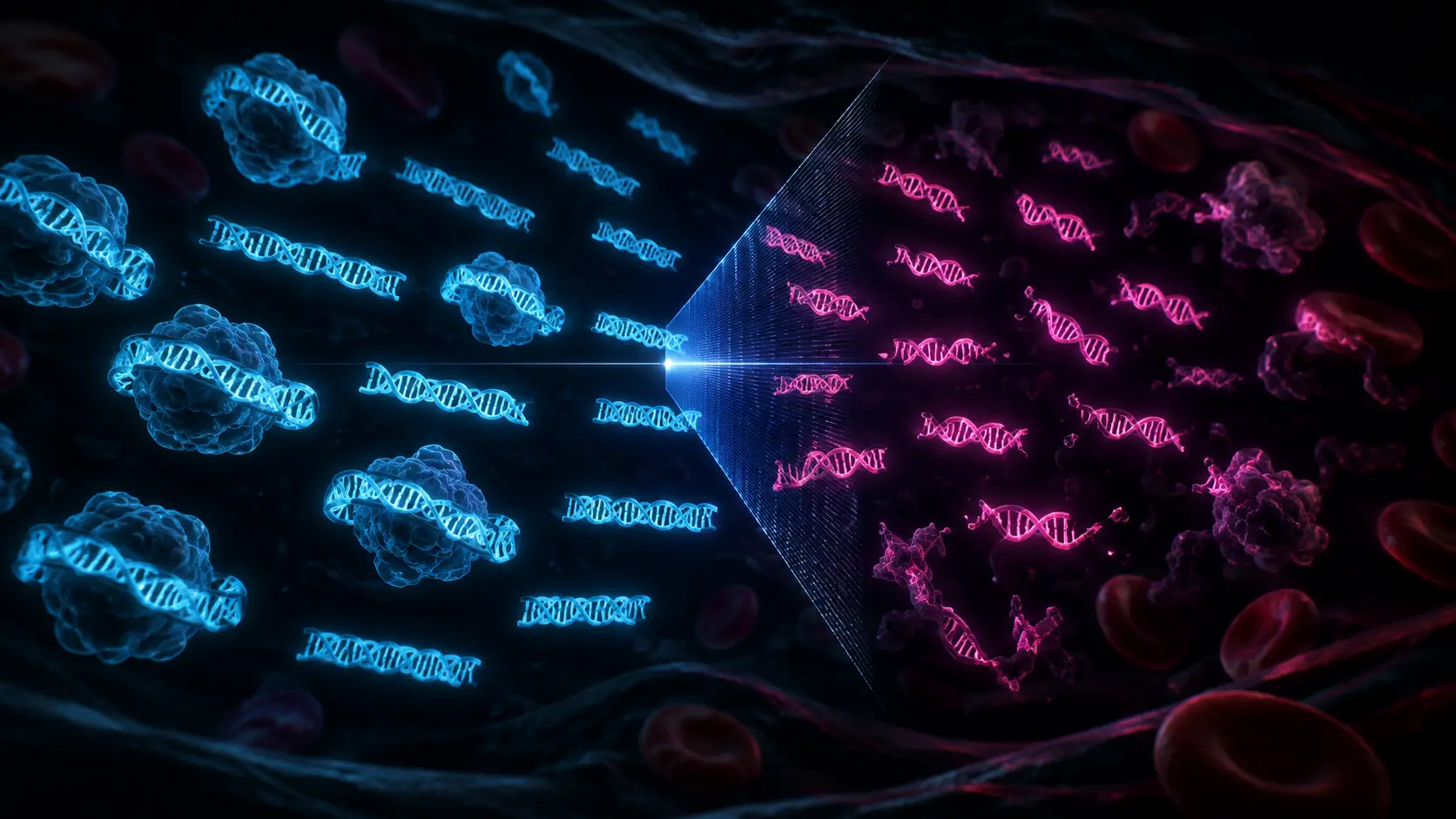

Imagine trying to find a single typo hidden inside a library of ten million books. That is the fundamental flaw of today’s most advanced cancer blood tests. Standard liquid biopsies operate on a simple premise: tumors shed pieces of their DNA into your bloodstream, and if a laboratory can sequence that DNA and find a specific genetic spelling mistake, they can diagnose the cancer. The problem is that early-stage tumors shed almost nothing. The microscopic trace of cancer DNA is entirely drowned out by the massive ocean of healthy DNA floating in your veins, causing traditional liquid biopsies to miss the most critical, early-stage warnings.
Why should you care right now? Because biotechnology has stopped looking for the typo and started looking at how the pages are torn. In a radical departure from traditional genetics, a breakthrough field known as “Fragmentomics” is moving beyond mutations. Scientists have discovered that when a cancer cell dies, its DNA breaks apart in a physically different shape and length than a healthy cell. By using machine learning to analyze the structural decay and “cut patterns” of millions of broken DNA fragments in a single vial of blood, oncologists are bypassing the sensitivity limits of legacy tests. This structural approach is finally making reliable, early-stage multi-cancer detection a clinical reality.
What is Fragmentomics?
Fragmentomics is an advanced field of genomic medicine that analyzes the physical size, cut patterns, and structural degradation of cell-free DNA (cfDNA) floating in the bloodstream. Instead of searching for specific genetic mutations, it evaluates the global structural profile of degraded DNA to identify the presence and tissue origin of cancer.
At a Glance
- Concept: Identifying cancer not by reading the genetic code, but by looking at the physical length and jagged edges of the broken DNA strings floating in a patient’s blood.
- Why it matters: Standard tests look for a specific mutation (a needle in a haystack). Fragmentomics looks at the entire haystack. It requires far less tumor DNA to trigger a positive result, dramatically improving early-stage cancer detection.
- Who uses it: Cutting-edge liquid biopsy developers (like Delfi Diagnostics), molecular oncologists, and pharmaceutical companies tracking minimal residual disease (MRD).
- Biggest takeaway: Because different organs package their DNA differently, the way the DNA breaks acts as a geographic barcode. Fragmentomics can not only tell a patient they have cancer, but exactly which organ it is growing in.
In Simple Words
Inside your cells, DNA is not a loose, tangled string; it is neatly wrapped around millions of tiny protein spools, much like thread wrapped around a wooden spool.
When a cell dies, it gets chopped up and thrown into the bloodstream. The biological “scissors” that do the chopping can only cut the string in the exposed spaces between the spools. The thread wrapped tightly around the spool is protected.
A healthy cell and a cancer cell use different parts of their DNA, meaning their “spools” are spaced differently. Therefore, when a healthy cell is chopped up, it produces pieces of thread that are all exactly 167 inches long. But when a chaotic cancer cell is chopped up, it produces highly irregular pieces that are much shorter, with jagged edges.
Fragmentomics simply scoops up all the thread floating in your blood and measures it with a ruler. If the computer sees too many short, jagged pieces of thread, it instantly flags the presence of cancer, even if it cannot read the actual genetic letters printed on the thread itself.
Why This Matters
For Biotech Investors, Oncologists, and Pharma Executives, fragmentomics solves the core economic and biological bottleneck of the Multi-Cancer Early Detection (MCED) market.
Ultra-deep targeted sequencing—the traditional method of searching for rare mutations—requires sequencing a small panel of genes thousands of times over to ensure accuracy. This is astronomically expensive and often yields false negatives if the patient’s specific tumor doesn’t happen to carry the exact mutations on the panel. Fragmentomics flips the economics. It utilizes low-coverage Whole Genome Sequencing (lcWGS). Because it only needs a shallow, wide scan of the genome to measure the lengths and endpoints of the fragments, the sequencing costs plummet. It transforms liquid biopsy from a bespoke, high-cost mutation hunt into a cheap, universal, scalable screening tool for global populations.
From Mutational ctDNA to Epigenetic Fragmentomics
Fragmentomics represents the transition from genetic sequencing to epigenetic profiling.
Genetics tells you the static code you were born with. Epigenetics tells you how that code is actually packaged, folded, and actively used by a living cell. By analyzing the physical degradation patterns of cell-free DNA (cfDNA), scientists are essentially reverse-engineering the epigenetic state of the dying cell. It is a paradigm shift: the “garbage” degradation patterns of dead DNA are actually a high-fidelity mirror reflecting the exact biological state of the hidden tumor.
How cfDNA Fragmentomics and Nucleosome Footprinting Work
Extracting clinical diagnoses from the chaos of degraded DNA requires mapping the mechanics of cellular apoptosis. Here is the first-principles breakdown of the architecture.
1. The Fundamental Problem: The Sensitivity Floor
In Stage I and Stage II cancers, the “tumor fraction” (the percentage of cfDNA in the blood that actually comes from the tumor) is frequently below 0.1%. Finding a mutated piece of DNA at this concentration is mathematically improbable, causing standard liquid biopsies to suffer from severe sensitivity floors.
2. The Core Mechanism: Nucleosome Footprinting
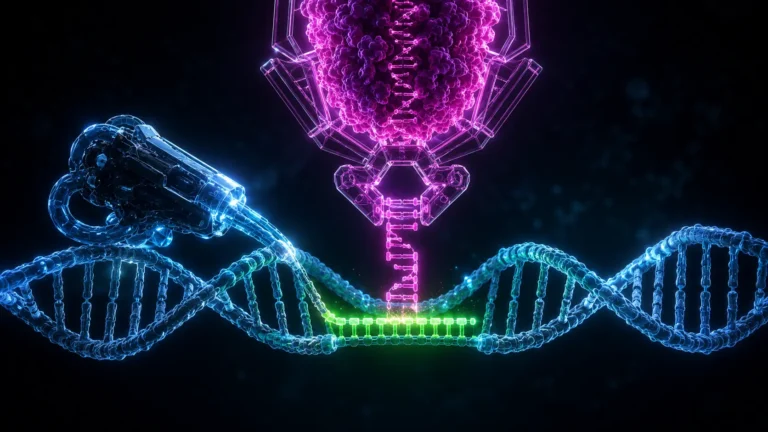
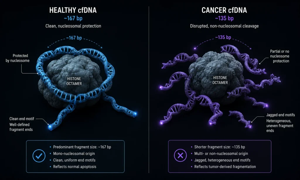
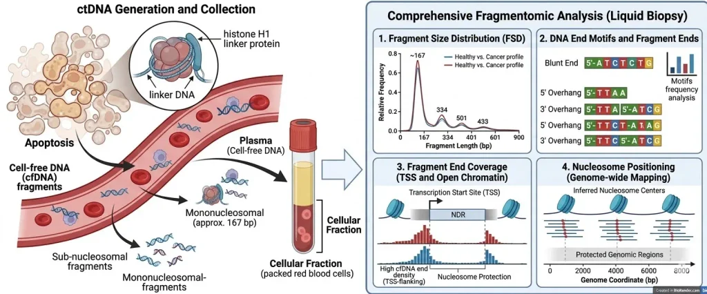
Human DNA is tightly coiled around histone protein complexes called nucleosomes. During apoptosis (programmed cell death), endonuclease enzymes cleave the DNA. They act like scissors, cutting the exposed linker DNA between the nucleosomes. Because the DNA wrapped around the nucleosome is protected from the scissors, the resulting fragments naturally measure roughly 167 base pairs (bp) in length.
3. Technical Depth: Fragment Size Distribution
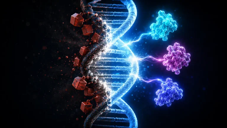
Cancer cells undergo massive chromosomal instability and chaotic gene expression. Their chromatin is disorganized, and they frequently die via necrosis (violent cell death) rather than orderly apoptosis. This chaotic death means the DNA is chopped up haphazardly.
When oncologists sequence the blood, they look at the Fragment Size Distribution. Healthy cfDNA creates a sharp peak at 167 bp. Cancer cfDNA creates an irregular, shifted profile, heavily burdened with abnormally short fragments (typically 130–145 bp).
4. Technical Depth: End Motifs and Jagged Ends
Beyond just length, the specific enzymes that cut the DNA leave behind signature chemical cuts, known as “end motifs” (the exact 4-letter sequence at the tip of the broken strand). Furthermore, cancer cfDNA frequently exhibits “jagged ends”—where the double helix is broken unevenly, leaving single-stranded overhangs. Measuring the frequency of these jagged ends provides an independent, highly sensitive variable for detecting malignancy.
5. Real-World Consequences: Machine Learning Classification
No human can manually review millions of fragmented DNA strands. The raw lcWGS data is fed directly into complex machine-learning algorithms. The AI evaluates the genome-wide fragmentation patterns, copy number variations (CNVs), and end motifs simultaneously. Because the algorithm evaluates the entire global footprint rather than a single mutated locus, it requires vastly less tumor DNA to establish a statistically significant cancer diagnosis.
Clinical Applications: MCED, MRD, and Tissue of Origin
Fragmentomics is rapidly transitioning from academic validation into the commercial diagnostic market, targeting the deadliest, fastest-moving cancers.
High-Grade Serous Ovarian Carcinoma (HGSOC): Ovarian cancer is notoriously difficult to detect because it lacks clear early symptoms and standard biomarkers (like CA-125) are notoriously unreliable. By the time it is caught, it is usually Stage III or IV. In clinical validation studies targeting ASCO 2026 guidelines, fragmentomic platforms demonstrated unprecedented ability to detect HGSOC in Stage I and II. By evaluating the chaotic nucleosome footprint of ovarian tissue, the test provides a highly sensitive, non-invasive triage tool for women presenting with ambiguous pelvic masses.
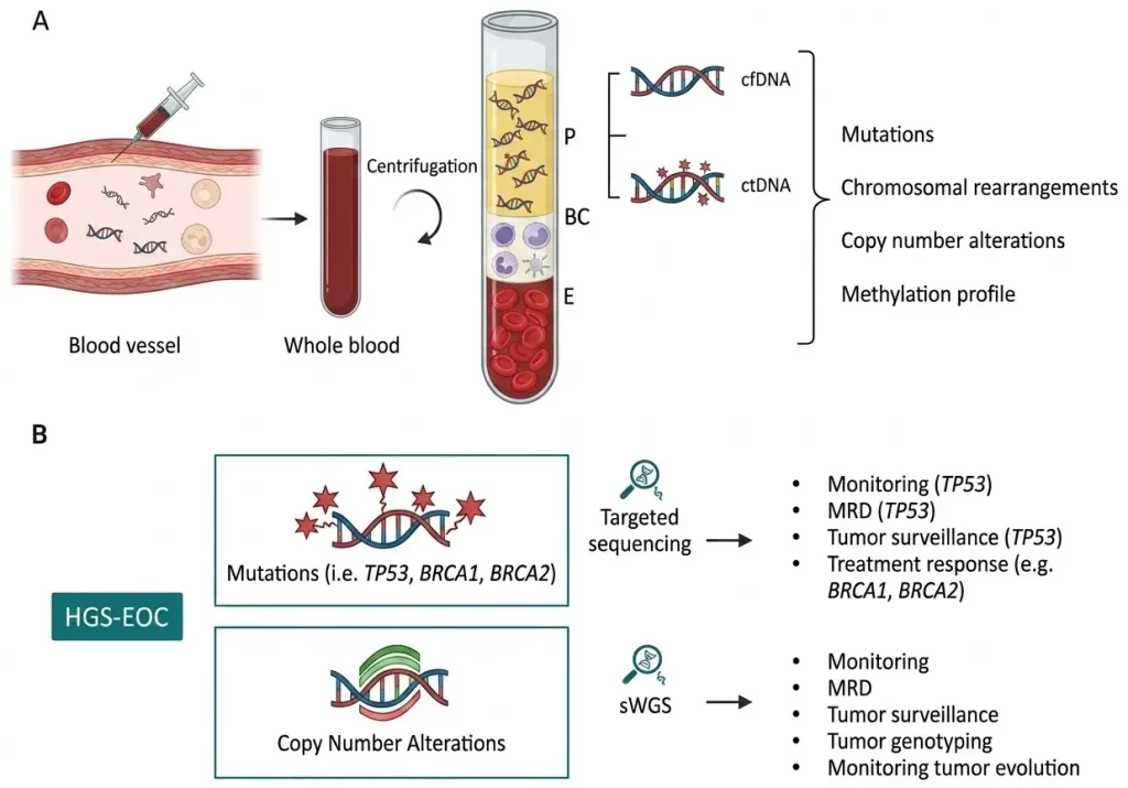
Minimal Residual Disease (MRD) Monitoring: After a surgeon removes a lung or colon tumor, the ultimate question is: Did we get it all? Traditionally, oncologists order a custom, highly expensive mutational panel based on the patient’s specific excised tumor to track if the cancer returns. Fragmentomics offers an “off-the-shelf” alternative. By simply tracking the global fragment size distribution in the patient’s blood over time, doctors can detect the microscopic structural changes that signal a cancer relapse months before a new tumor appears on a CT scan, without needing to customize the test for each individual patient.
Tissue of Origin (TOO) Mapping: Finding cancer early is only helpful if the doctor knows where to look for it. Because different organs have different active genes, the nucleosome spacing in liver tissue is physically different than in breast tissue. Fragmentomic AI models compare the broken DNA patterns in the blood against a master atlas of human chromatin accessibility. The test can not only flag the presence of a tumor but output a predictive heat map, telling the oncologist to immediately order an MRI of the pancreas rather than initiating a blind, full-body hunt.
Economic & Strategic Impact
The core economic disruption of Fragmentomics is the Democratization of Sequencing Costs.
Traditional mutational liquid biopsies rely on “Ultra-Deep Targeted Sequencing.” To find a rare mutation, the sequencing machine must read the same targeted gene 10,000 times to ensure the mutation isn’t just a machine error. This depth requires massive, expensive sequencing flow cells, keeping the cost of clinical liquid biopsies artificially high (often exceeding $3,000 per test).
Fragmentomics relies on low-coverage Whole Genome Sequencing (lcWGS). Because it is evaluating the macroscopic pattern of the entire genome rather than hunting for a microscopic needle, it only needs to read the genome 1 to 2 times over (1x – 2x depth). This reduces the raw sequencing cost to a fraction of traditional methods. As these costs collapse, liquid biopsy transitions from a specialized, late-stage oncology tool into a cheap, routine screening deployed in standard primary care physicals.
Advantages
- Universal Applicability: Standard tests only work if the patient’s tumor happens to possess the specific mutation the test is looking for. Fragmentomics requires no prior knowledge of the tumor’s genetics; it detects the universal structural chaos of cancer cell death.
- Superior Early-Stage Sensitivity: By evaluating millions of fragments across the entire genome rather than a handful of targeted loci, fragmentomics achieves a mathematically superior “signal-to-noise” ratio in low-shedding Stage I and II cancers.
- Lower Sequencing Costs: The reliance on low-coverage sequencing heavily reduces laboratory overhead, allowing diagnostic companies to scale production and expand profit margins while lowering the retail cost of the test.
- Tissue of Origin Identification: Captures the epigenetic signature of the DNA, allowing algorithms to accurately predict exactly which organ the tumor is originating from.
Limitations
- Pre-Analytical Vulnerabilities: Fragmentomics relies entirely on the physical size of the DNA. If a phlebotomist shakes the blood tube too aggressively, or if the sample sits in a warm truck for two days, the healthy DNA physically degrades and snaps into shorter pieces. This artificial degradation mimics the cancer signature, triggering a devastating false positive.
- Massive AI Training Data Requirements: The algorithms must be trained to differentiate between the nucleosome footprint of cancer and the footprint of benign inflammatory diseases (like rheumatoid arthritis or hepatitis). Gathering massive, clinically annotated datasets to train the AI against these confounding variables is agonizingly slow.
- Biological Confounders: Pregnancy, intense exercise, and severe infections all trigger massive cell death and release altered cfDNA fragments into the blood, potentially confusing the diagnostic algorithm.
Common Misconceptions
Misconception: Fragmentomics replaces genetic sequencing completely.
Reality: Fragmentomics detects that the cancer is there, and where it is. But to determine exactly which targeted drug (like a specific kinase inhibitor) will cure it, the oncologist still needs a traditional mutational liquid biopsy to read the specific genetic code of the tumor.
Misconception: The test physically extracts the cancer cells from the blood.
Reality: The test extracts cell-free DNA (cfDNA)—dead, broken molecules floating in the plasma. It does not extract Circulating Tumor Cells (CTCs), which are intact, living cancer cells.
Misconception: Short DNA always means cancer.
Reality: While cancer cfDNA is typically shorter, mitochondrial DNA and cfDNA from certain healthy tissues are also naturally short. The diagnosis relies on the global distribution and machine-learning pattern recognition, not just a binary “short equals cancer” rule.
What Most People Miss
The disruptive intelligence value of Copy Number Alterations (CNAs) at Low Coverage.
Most reporting on fragmentomics focuses exclusively on the length of the fragments. What most miss is that lcWGS simultaneously captures Copy Number Alterations for free.
As cancer ravages a cell, it often duplicates entire sections of chromosomes (amplifications) or deletes them. Because fragmentomics already scatters sequencing reads across the entire genome to measure fragment length, the algorithm inherently sees if there is a massive overrepresentation or underrepresentation of DNA from a specific chromosome. By mathematically fusing fragment size distribution with Copy Number Alteration data, the AI creates an interlocking, dual-layered diagnostic matrix that drastically reduces the false positive rate.
Comparison Table
| Feature | Solid Tissue Biopsy | Mutational ctDNA (Targeted NGS) | Fragmentomics (lcWGS) |
| Analyte Targeted | Intact tumor cells | Specific genetic mutations | Structural DNA degradation patterns |
| Invasiveness | High (Surgical) | Low (Blood draw) | Low (Blood draw) |
| Sequencing Depth | Low to Moderate | Ultra-Deep (>5,000x) | Ultra-Low (1x – 2x) |
| Early-Stage Sensitivity | Excellent (if found) | Poor to Moderate | High |
| Universal Detection | N/A | No (Panel dependent) | Yes (Agnostic to mutations) |
Case Study
Situation: The clinical reality of High-Grade Serous Ovarian Carcinoma (HGSOC) is grim; the vast majority of patients are diagnosed at advanced stages, resulting in a 5-year survival rate below 30%. Early-stage HGSOC sheds very little mutated ctDNA, rendering traditional mutation-hunting liquid biopsies functionally useless for early screening.
Challenge: Develop a highly sensitive, non-invasive blood test capable of detecting the faint epigenetic footprint of Stage I/II HGSOC, while maintaining a low false-positive rate to avoid triggering unnecessary prophylactic surgeries in healthy women.
Solution (The DELFI Validation Pathway): Researchers utilizing the DELFI (DNA evaluation of fragments for early interception) methodology focused on the genome-wide fragmentation profiles of women with adnexal masses. Instead of looking for BRCA or TP53 mutations, they sequenced the cfDNA at 1x to 2x depth and fed the fragment size distributions, end motifs, and copy number variations into a machine-learning classifier trained specifically on the nucleosome packaging of ovarian tissue.
Outcome: The fragmentomic classifier achieved a mathematically profound separation between malignant HGSOC and benign masses. In clinical validation cohorts leading into 2026 guidelines, the structural analysis demonstrated sensitivity rates exceeding 80% for early-stage disease, vastly outperforming legacy CA-125 protein biomarkers and targeted mutational panels.
Lessons Learned: The deployment validated the core thesis of fragmentomics: the structure of cell death is a louder alarm bell than the genetic code of the tumor. By shifting the computational burden from deep sequencing hardware to advanced machine-learning software, the industry secured the blueprint for expanding Multi-Cancer Early Detection into the deadliest, most stealthy oncological verticals.
Future Outlook
Next 12–24 Months
The era of Symptomatic Triage and MRD dominance. The immediate commercial rollout of fragmentomics will be carefully constrained to high-prevalence cohorts. Instead of testing the general public, oncologists will use fragmentomic tests for “symptomatic triage”—for example, testing a patient who has a suspicious nodule on a lung CT scan to determine if an invasive surgical biopsy is necessary. Simultaneously, the low cost of lcWGS will make fragmentomics the default technology for Minimal Residual Disease (MRD) monitoring, allowing doctors to test post-operative cancer patients every 30 days to catch recurrences instantly.
Next 3–5 Years
The scaling of Multiomics and Methylation Fusion. The absolute limit of analyzing purely broken DNA fragments will be reached. To push sensitivity past 95%, diagnostic companies will fuse fragmentomics with epigenetic methylation profiling (analyzing the chemical “on/off” tags attached to the DNA). This “Multiomic” approach will require entirely new classes of deep learning neural networks capable of ingesting fragment size, end motifs, copy number variations, and methylation density simultaneously. This resulting super-algorithm will become the standard engine for comprehensive Multi-Cancer Early Detection (MCED) platforms.
Next 10 Years
The Universal Primary Care Integration. By the mid-2030s, the cost of low-coverage Whole Genome Sequencing will fall to negligible levels. Fragmentomic liquid biopsies will cease to be specialized oncological tools; they will be integrated into the standard annual blood panel drawn during a routine physical, alongside cholesterol and glucose checks. By shifting the global healthcare model from reactive surgical intervention to proactive, asymptomatic interception, fragmentomics will structurally lower global cancer mortality rates, fundamentally redefining the economics of health insurance and life extension.
Most Likely Scenario
Fragmentomics is the definitive, thermodynamic solution to the biological noise of the human bloodstream. By accepting that early-stage tumors do not shed enough specific mutations to be caught reliably, the industry has executed a brilliant pivot toward structural pattern recognition. Driven by the exponentially dropping cost of shallow sequencing and the relentless advancement of AI classifiers, fragmentomics will secure its position as the foundational architecture of 21st-century cancer diagnostics.
Key Takeaways
- Standard liquid biopsies try to detect cancer by hunting for specific genetic mutations (spelling mistakes) in the blood. They often fail because early-stage cancers shed very little DNA.
- Fragmentomics analyzes the physical size, shape, and cut patterns of broken DNA strings instead. It looks at the “garbage” left behind when a cell dies.
- Because cancer cells package their DNA differently than healthy cells (altering the spacing of their “nucleosome” spools), they break into distinct, shorter, jagged fragments.
- The test uses machine learning to scan the entire genome at a shallow depth, finding the chaotic structural footprint of cancer far earlier than mutation-hunting tests.
- Because it doesn’t need to read genes thousands of times over to find a single mutation, fragmentomics relies on low-coverage sequencing, making the test vastly cheaper and more scalable.
- The technology is heavily focused on detecting deadly, stealthy cancers like ovarian cancer (HGSOC) and tracking if a tumor has returned after surgery (MRD).
Glossary
Apoptosis: The orderly, programmed death of a cell. When a cell undergoes apoptosis, specialized enzymes cut its DNA into neat, predictable lengths.
Cell-Free DNA (cfDNA): Fragments of dead, broken DNA floating freely in the blood plasma, shed by both healthy cells and tumors.
Copy Number Alteration (CNA): A type of genetic mutation where entire sections of a chromosome are duplicated or deleted, leading to an abnormal amount of DNA from a specific region.
End Motif: The specific sequence of 4 to 6 genetic letters at the exact tip of a broken DNA fragment. Cancer cfDNA often exhibits distinct, jagged end motifs.
Low-Coverage Whole Genome Sequencing (lcWGS): A fast, cheap method of sequencing where the machine only reads across the entire genome 1 or 2 times, gathering a broad overview rather than deep, specific details.
Nucleosome: A tiny protein spool (made of histones) that DNA wraps tightly around for protection and packaging inside the cell.
Sources
Nature: Fragmentation patterns of cell-free DNA
Science Translational Medicine: Genome-wide cell-free DNA fragmentation in patients with cancer (DELFI)
American Society of Clinical Oncology (ASCO): Liquid Biopsy for Early Detection of High-Grade Serous Ovarian Carcinoma
Cell: Nucleosome footprinting in cell-free DNA
Delfi Diagnostics: DELFI Platform and Fragmentomics Overview