Advertisement
)
AT A GLANCE
- Concept: Vitrification: Liquid ethane flash-freezes biological samples so rapidly that water cannot form destructive ice crystals.
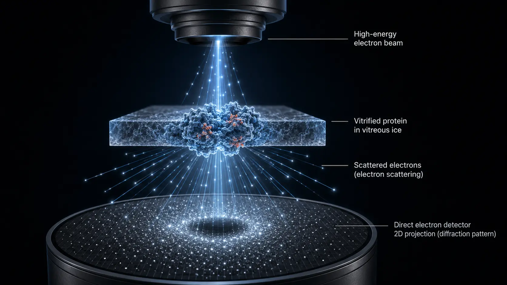
- Concept: Electron Scattering: Electrons pass through the frozen protein, deflecting off its atomic nuclei to cast a two-dimensional shadow.
- Concept: Single-Particle Analysis: Algorithms compile millions of two-dimensional shadows from different angles into a three-dimensional atomic model.
- Concept: AI Foundation: These exact physical coordinates provide the mandatory training data that powers artificial intelligence folding algorithms.
HOW IT WORKS
Biological proteins are highly unstable, folding and unfolding constantly in liquid environments. Trying to image them with traditional light microscopes fails due to strict optical diffraction limits, while legacy X-ray crystallography requires forcing them into unnatural, rigid crystal structures. Cryogenic electron microscopy (Cryo-EM) solves this physical limitation by executing a sub-millisecond freeze-frame.
Technicians plunge a purified protein solution into liquid ethane cooled to -180°C. This extreme thermal drop occurs so fast that the water molecules freeze in place without forming jagged ice crystals. The process suspends the protein in a glass-like state called vitreous ice, perfectly preserving its exact native shape.
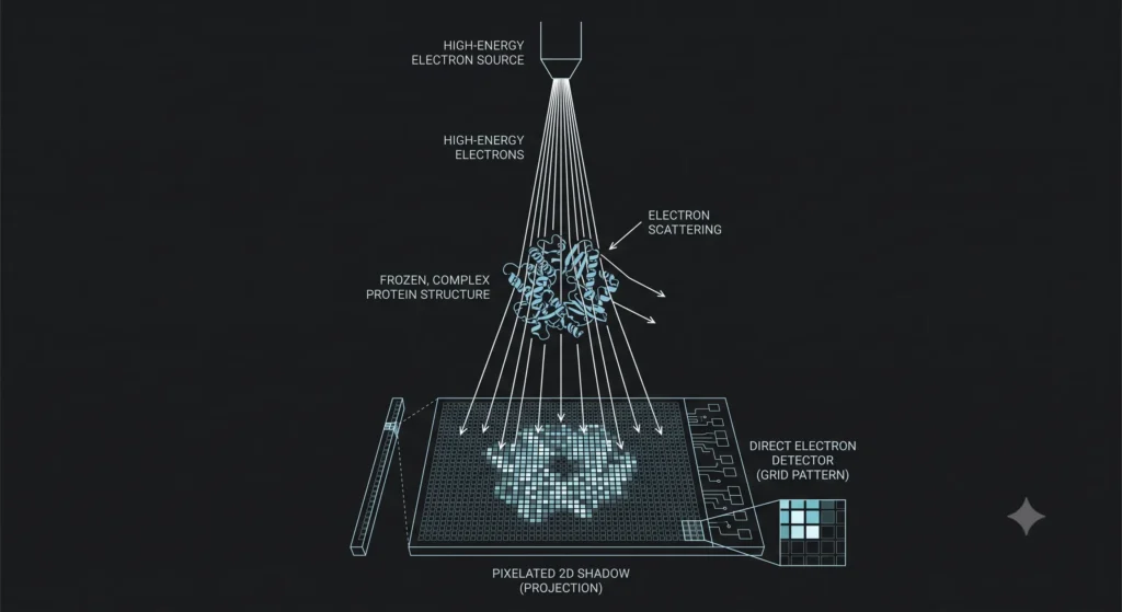
Inside a massive vacuum chamber, an electron gun fires a concentrated beam through this vitrified sample at near light speed. The ability to resolve individual atoms relies on the extreme kinetic energy of the beam, dictated by the de Broglie wavelength equation:
$$\lambda = \frac{h}{\sqrt{2 m_e e V}}$$
Where λ (lambda) is the electron wavelength, h is Planck’s constant, m_e is the electron mass, e is the elementary charge, and V is the accelerating voltage. Operating at 300 kilovolts generates an electron wavelength significantly smaller than the diameter of a single carbon atom.
As these high-speed electrons pass through the frozen protein, they collide with its atomic nuclei and scatter. A direct electron detector sitting at the base of the machine intercepts these scattered particles. This semiconductor sensor records the exact location of every single electron strike, creating a highly detailed, two-dimensional shadow of the protein.
The physical sample contains millions of identical proteins resting in random physical orientations. Advanced computational algorithms take thousands of these overlapping two-dimensional shadows and mathematically reverse-engineer them. The software compiles the varied angles into a flawless, three-dimensional topographical map of the entire target molecule.
WHY IT MATTERS NOW
The global pharmaceutical industry currently operates on the economics of rational drug design. Instead of mixing random chemicals and observing the biological result, scientists map a disease-causing protein and custom-build a synthetic molecule to physically block it. This precise manufacturing pipeline requires absolute structural certainty.
A single misplaced atom in a protein model causes a billion-dollar drug candidate to bind incorrectly, automatically failing late-stage clinical trials. Cryo-EM arrays provide the exact three-dimensional topography of highly complex, previously unmappable targets. These targets include human cell surface receptors and erratic viral spike proteins.
When the SARS-CoV-2 pathogen emerged, researchers used Cryo-EM to map the exact physical structure of its spike protein within a matter of weeks. This immediate physical blueprint allowed biomanufacturers to engineer synthetic mRNA vaccines that perfectly matched the viral docking mechanism. This speed physically accelerated the global biodefense response by years.
Operating these machines requires massive corporate or state-level capital expenditure. A top-tier Thermo Fisher Krios microscope costs upwards of ten million dollars and requires a dedicated, vibration-proof concrete bunker to isolate the sensitive electron beam from passing environmental traffic. This heavy capital requirement restricts high-level structural biology to elite research institutions and well-funded pharmaceutical conglomerates.
Consequently, controlling the physical hardware of structural biology dictates national pharmaceutical sovereignty. Nations that lack domestic Cryo-EM arrays cannot independently design modern precision therapeutics. They remain entirely dependent on licensing synthetic chemical blueprints from foreign biotech monopolies.
WHAT MOST PEOPLE MISS
Technology media attributes the recent leap in structural biology entirely to artificial intelligence, heavily praising software algorithms like DeepMind’s AlphaFold. Observers treat these algorithmic tools as independent scientific agents capable of replacing physical laboratories entirely. They view molecular mapping purely as a computational math problem.
They miss the absolute physical data dependency of the software layer. Artificial intelligence does not invent structural biological rules from thin air; it learned how proteins fold by ingesting the exact physical coordinates meticulously gathered by decades of X-ray and Cryo-EM hardware operations. Without the continuous stream of physical atomic verification produced by these massive electron microscopes, AI prediction models rapidly hallucinate and fail against novel biological variations.
THE TRAJECTORY
Next 12–36 Months: Cloud-based computational platforms will directly integrate with hardware arrays, processing electron scattering data in real-time. This integration will generate basic three-dimensional models while the physical sample remains inside the vacuum chamber, drastically accelerating operational throughput.
Next Five Years: Hardware manufacturers will commercialize time-resolved Cryo-EM. By firing microsecond lasers to unfreeze and refreeze samples instantly, researchers will capture proteins in motion, mapping the exact thermodynamic pathways of drug binding.
Next Ten Years: Cryo-electron tomography will map entire human cells in a single unbroken scan. Scientists will stop studying isolated proteins in purified solutions and begin mapping complex molecular interactions directly inside living tissue architecture.
What Could Go Wrong: The extreme volume of data generated—terabytes per day per machine creates a severe digital bottleneck. If storage and local transmission infrastructure fails to scale, research facilities will be forced to delete raw scattering data, permanently erasing the forensic evidence required to verify AI folding predictions.
Most Likely Outcome: The Cryo-EM array will become the mandatory physical anchor of the generative biology sector. AI will predict the vast majority of standard molecular structures, but high-end pharmaceutical development will always require physical electron scattering to verify and legally patent the final drug design.
KEY TERMS
- Cryogenic Electron Microscopy (Cryo-EM): An imaging technique that fires electrons through a flash-frozen biological sample to determine its exact molecular structure.
- Vitrification: The rapid cooling of a liquid medium into a glass-like state, preventing the formation of crystalline ice that would physically tear biological cells apart.
- Single-Particle Analysis: A computational method that combines millions of two-dimensional projection images of randomly oriented proteins into a single three-dimensional structural model.
- Direct Electron Detector: A highly sensitive semiconductor sensor that records individual electron strikes with extreme precision, replacing traditional optical camera lenses.
- Rational Drug Design: The invention of highly specific pharmaceuticals based on the known three-dimensional physical structure of the target biological molecule.
SOURCES
- Nature Methods — Single-Particle Cryo-Electron Microscopy at Atomic Resolution
- Thermo Fisher Scientific — Titan Krios and Direct Electron Detection Architecture
- National Institutes of Health (NIH) — The Impact of Cryo-EM on Structural Biology and Drug Discovery
- Journal of Molecular Biology — Time-Resolved Cryo-Electron Microscopy and Conformational Dynamics